Figure 6.

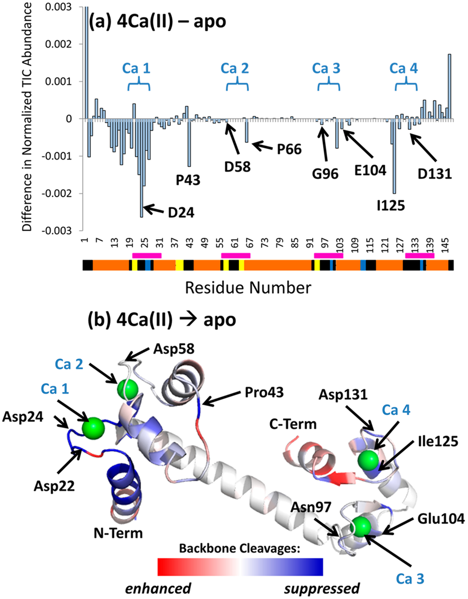
(a) Difference plot for the UVPD backbone cleavage yields of apo-calmodulin and the Ca(II) complexes (summed holo and apo fragment ions) (7+). Negative values represent regions of the protein backbone for which backbone cleavages of the metal complexes are suppressed relative to the apoprotein. The segmented bar beneath the residue numbers displays secondary structural elements: yellow (turn), blue (beta strand), orange (α helix), and black (unassigned). The binding sites for the four calcium ions are demarcated by pink bars. (b) The values from the difference plots are superimposed on the crystal structure (PDB: 1CLL) as heat maps to show areas of enhancement (red) and suppression (blue) of fragmentation upon metal binding. Selected residues are labeled for clarity. An expanded region of the Ca 1 binding site illustrating the polar contacts between the metal ion and the protein (side chains) is shown in Figure S21.
