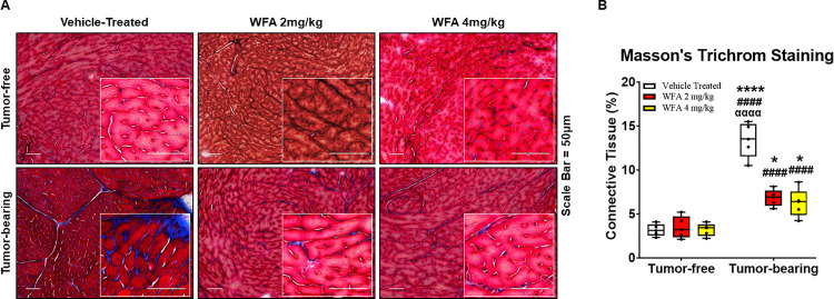
Fig 4. Withaferin A diminishes fibrotic scarring in the heart.
(A) Representative 20x images of Masson’s trichrome-stained midventricular heart sections in tumor-free and tumor-bearing mice that were treated with WFA or vehicle. Inset images are magnified from the displayed field of view. Scale bar = 50 μm. (B) Quantification of average collagen deposition. N = 10 per group. Black circles indicate individual data points. *p < 0.05; **p < 0.01; ***p < 0.001; or ****p < 0.0001 indicates a significant difference from the corresponding value of the tumor-free vehicle-treated group by two-way ANOVA followed by Tukey’s multiple comparison test. #p < 0.05 indicates a significant difference from the corresponding value of the tumor-free WFA 2 mg/kg group. αp < 0.05 indicates a significant difference from the corresponding value of the tumor-free WFA 4 mg/kg group. ¥p < 0.05 indicates a significant difference from the corresponding value of the tumor-bearing vehicle-treated group.

