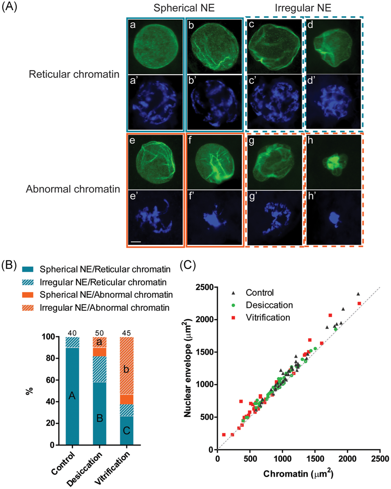
FIGURE 1.

Nuclear envelope conformation and chromatin configuration in cat germinal vesicles after desiccation and vitrification. (A) Representative micrographs of different categories of the nuclear envelope (NE) conformation (immunostaining of lamin A/C; a–h) and chromatin configurations (DAPI staining; a’–h’). Scale bar = 10 μm. (B) Proportion of different categories of nuclear envelope conformation and chromatin configuration in different treatment groups. Proportions with different letters (spherical NE/reticular chromatin category: capital letters; irregular NE/abnormal chromatin category: lowercase letters) differ within the category (p < .05). Numbers on the top of the bars indicate a total number of oocytes in each treatment group. (C) Areas of nuclear envelope relative to the area of the in oocytes after different treatments. The dotted line represents 1:1 ratio in which the size of chromatin equals that of the nuclear envelope
