Figure 4.

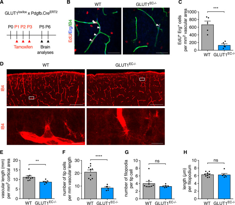
Loss of EC-GLUT1 (endothelial glucose transporter isoform 1) impairs neonatal brain angiogenesis. A, Schematic representation of experimental timing for brain analyses in GLUT1lox/lox×Pdgfb.CreERT2 pups. B and C, Representative pictures (B) and quantification (C) of 5-ethynyl-2’-deoxyuridine (EdU+)/ETS-related gene (Erg+) cells in the primary plexus of P5 GLUT1EC−/− pups vs wild-type (WT) littermates (Student t test). White arrows (B) indicate EdU+/Erg+ cells. D and H, Representative pictures showing the cortical area and a tip cell magnification of IB4 (isolectin griffonia simplicifolia B4)-stained thick brain sections from P6 GLUT1EC−/− pups vs WT littermates (D) and corresponding quantifications of vascular length (E) tip cell number (F) filopodia number per tip cell (G) and filopodial length (H) (Student t test or Mann-Whitney U test). Scale bar=50 µm (B), 200 µm for upper and 10 µm for lower (D). **P<0.01, ***P<0.001, and ****P<0.0001.
