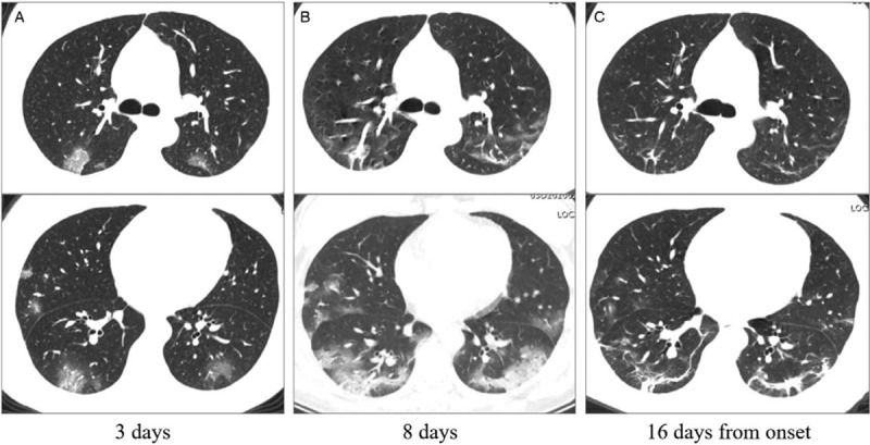
Figure 3.

The initial computed tomography (CT), CT with most obvious opacities, and CT with decreased opacities of a 38 years old nurse. A: Patchy ground-glass opacities distributed bilaterally on the initial CT (3 days from onset). B: In the follow-up CT, ground-glass opacities progressed to multiple ground-glass infiltration in the lungs. C: Fibrous stripes could be a common sign during the remission stage. Images in the first and second lines represent different cross sections.
