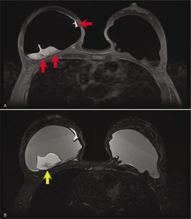
Figure 3.

(A) Breast MR confirms the presence of a large periprosthetic seroma visible on the right sinus (red arrows). (B) In the corresponding T2-mode MR, amorphous material adhered to the back of the right prosthesis and due to fibrin deposits (yellow arrow) is recognizable.
