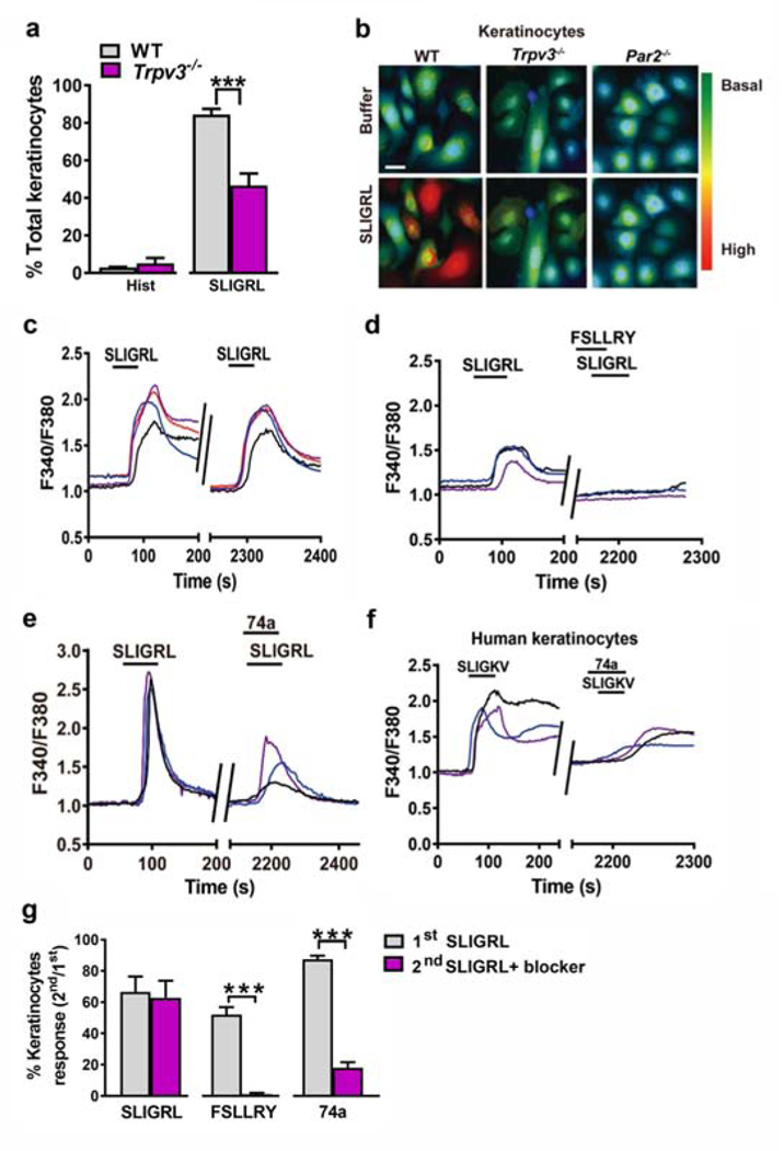
Figure 2. TRPV3 is involved in PAR2/Ca2+ signaling in keratinocytes.
(a) The percentages of responding keratinocytes from WT and Trpv3−/− mice stimulated with histamine (100 μM) and SLIGRL (50 μM). ***p < 0.001, unpaired t tests. n = 3 mice per group. (b) Representative fluorescence images of Fura2 (2 μM)-loaded WT, Trpv3−/− and Par2−/− keratinocytes stimulated with SLIGRL (50 μM). Scale bar, 20 μm. (c) Representative traces showing intracellular Ca2+ responses elicited by SLIGRL (50 μM) in WT mouse keratinocytes. (d, e) The effect of FSLLRY (100 μM) (d) and 74a (100 μM) (e) on SLIGRL-induced Ca2+ responses in WT mouse keratinocytes. (f) The effect of 74a (100 μM) on SLIGKV-induced (100 μM) Ca2+responses in human keratinocytes. (g) Quantified data showing the percentages of SLIGRL-responsive keratinocytes before and after the incubation of FSLLRY or 74a. ***p < 0.001, unpaired t test, n = 3 mice per group. Data are presented as mean ± s.e.m.

