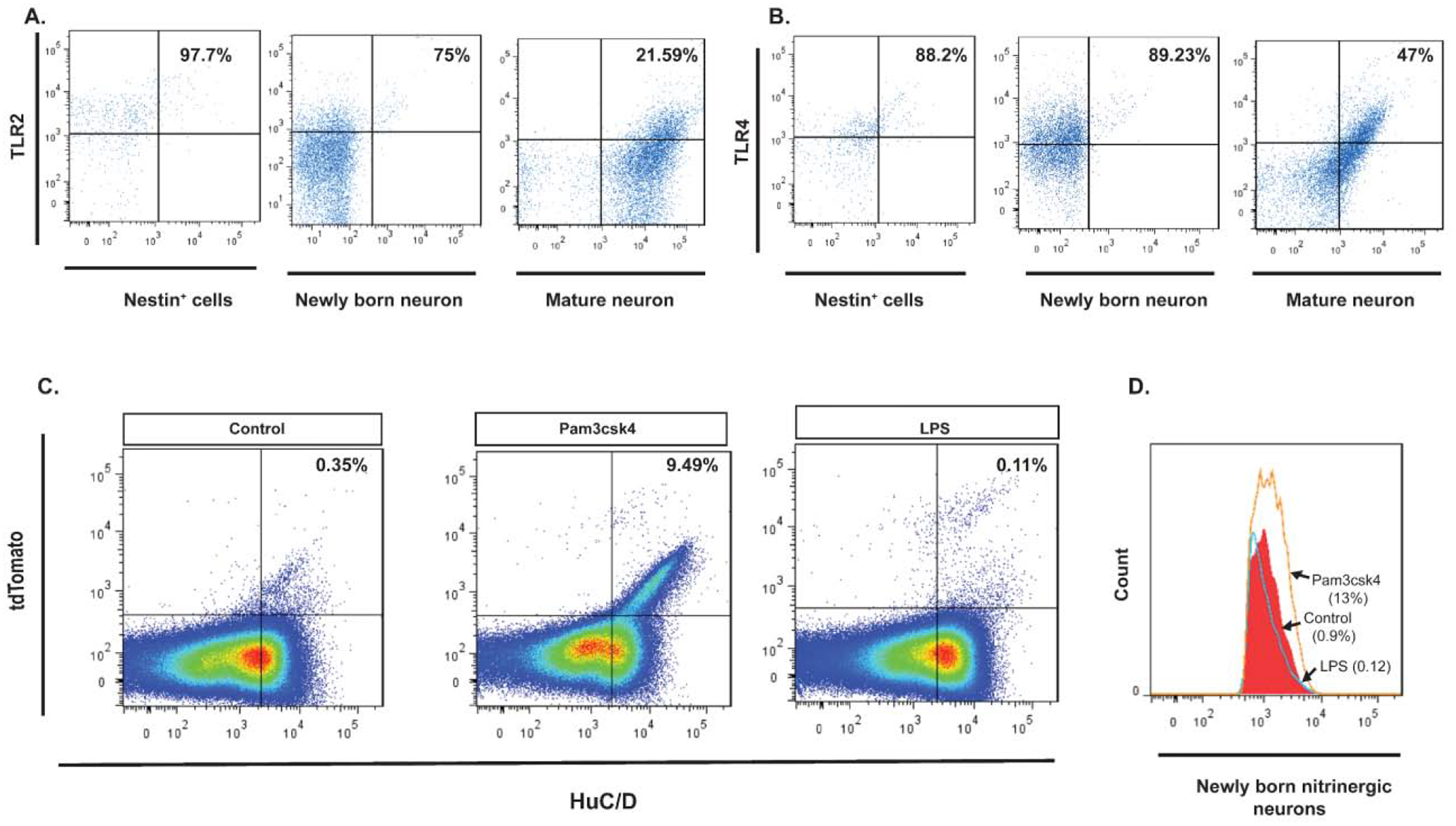
Figure 1. Expression of TLR2 and TLR4 receptors by Nestin+ ENPCs and the effects of their activation.

(A. left panel) Flow cytometry of Nestin+ ENPCs isolated from colonic myenteric plexus of Nestin-GFP mice and stained with TLR2, showing that 97.7% of Nestin+ cells express TLR2 (top right quadrant). (A. middle panel) Flow cytometry of Huc/D+ isolated from colonic myenteric plexus of Nestin-creERT2:tdTomato mice after tamoxifen induction and stained with TLR2, showing that 75% of HuC/D+tdTomato+ neurons express TLR2. (A. right panel) Flow cytometry of HuC/D+ neurons that do not express tdTomato shows that 21.59% of these cells express TLR2. (B. left panel) Flow cytometry of Nestin+ ENPCs isolated from colonic myenteric plexus of Nestin-GFP mice and stained with TLR4, showing that 88.2% of Nestin+ cells express TLR4. (B. middle panel) Flow cytometry of Huc/D+ isolated from colonic myenteric plexus of Nestin-creERT2:tdTomato mice after tamoxifen induction and stained with TLR4, showing that 89.2% of HuC/D+tdTomato+ neurons express TLR4. (B. right panel) Flow cytometry of HuC/D+ neurons that do not express tdTomato shows that 47% of these cells express TLR4. (C) Pseudocolor plots of flow cytometry data of cells isolated from colonic myenteric plexus of Nestin-creERT2:tdTomato mice after tamoxifen induction and being cultured for 14 days and stained for HuC/D. (C. left panel) Flow cytometry from control culture group showing newly formed neurons from Nestin+ ENPCs that express tdTomato (top right quadrant). (C. middle panel) Addition of TLR2 agonist (Pam3csk4) increased the number of newly formed neurons. (C. right panel) Addition of TLR4 agonist (LPS) did not increase the population of newly formed neurons. (D) Flow cytometry of HuC/D+nNOS+ cells isolated from colonic myenteric plexus of Nestin-creERT2:tdTomato mice and cultured for 14 days with addition of Pam3csk4 (orange line), LPS (blue line), or vehicle (red filled curve) showing increase in the population of newly formed nitrergic neurons with TLR2 agonist addition.
