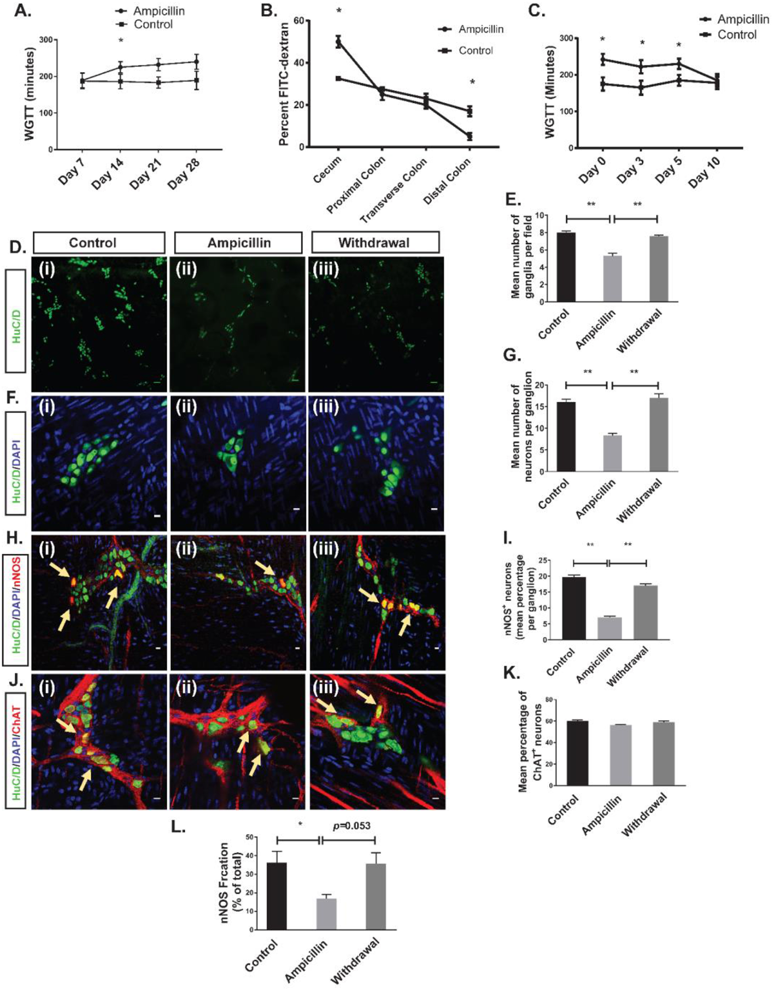
Figure 3. Effects of ampicillin on colon transit time and myenteric neurons.

(A) Ampicillin prolonged whole gut transit time after 2, 3, and 4 weeks of treatment. (B) FITC dextran test showed that the slowing of transit time occurs predominantly in the cecum. (C) WGTT normalized 10 days after discontinuation of ampicillin. (*p<0.05). D (i-iii) and F (i-iii) are representative photomicrographs of colonic myenteric plexus after immunostaining with the pan-neuronal marker HuC/D (green); nuclei are stained with DAPI (blue). H (i-iii) are representative photomicrographs of a colonic myenteric ganglion after immunostaining with the neuronal marker HuC/D (green) and nitrergic neuron marker nNOS (red); nuclei are stained with DAPI (blue).J (i-iii) are representative images of the colon wall from a ChAT-cre:tdTomato mouse where myenteric cholinergic neurons express red fluorescent protein tdTomato (red) after immunostaining with the neuronal marker HuC/D. Nuclei are counter stained with DAPI. (D (i)) Immunostaining with antisera against HuC/D (green) of myenteric plexus in control mice shows normal ganglion density. (D (ii)) Ampicillin treatment reduced the ganglion density. (D (iii)) Ganglion density recovered 10 days after discontinuation of ampicillin. (E) Grouped results of ganglion density under various conditions (**p<0.001). (F (i)) Immunostaining with antisera against HuC/D shows neurons in a representative colonic myenteric ganglion in control mice. (F (ii)) Ampicillin treatment reduced the number of neurons per ganglion. (F (iii)) 10 days after discontinuation of ampicillin, the number of neurons per ganglion normalized. (G) Grouped results of neuronal number per ganglion (** p<0.001). (H (i)) Representative image of a normal myenteric ganglion including nitrergic neurons (arrow). (H (ii)) Ampicillin treatment reduced the number and percentage of nitrergic neurons (H (iii)) Number of nitrergic neurons (arrows) and percentage of nitrergic neurons recovered 10 days after discontinuation of ampicillin. (I) Grouped results of percentage of nitrergic neurons per ganglion under various conditions (*p<0.05). (J (i)) Representative image of a normal myenteric ganglion including cholinergic neurons (arrow). (J (ii)) Ampicillin treatment did not reduce the percentage of cholinergic neurons (J (iii)) Percentage of cholinergic neurons did not change 10 days after discontinuation of ampicillin. (K) Grouped results of percentage of cholinergic neurons per ganglion under various conditions (p=0.31). (L) Mean nNOS percentage of the total NO released by the LM-MP isolated from the cecum in three different groups of control, ampicillin and withdrawal (*P < 0.01). Scale bar is 100 μm in D panel and 10 μm in F, H and J panel.
