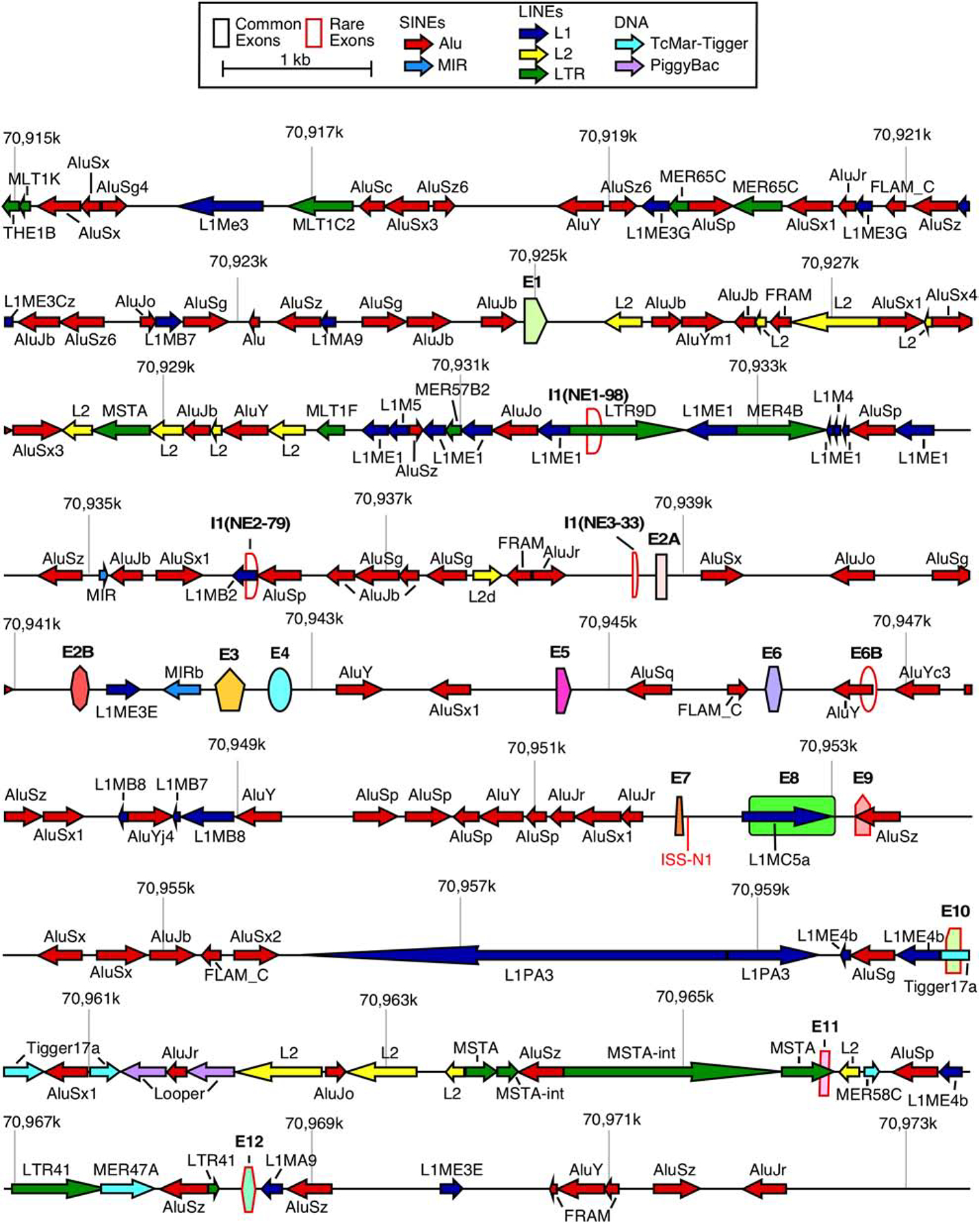
Figure 1. Organization of the SMN genes.

A scale depiction of the SMN1 gene (SMN2 has an identical overall structure) and flanking sequences are shown. Exons are depicted by colored shapes. Common exons (≥10% of total SMN RNA) are outlined in black, rare exons (<10% of total SMN RNA) are outlined in red. Repeat sequences as identified by Repeatmasker are depicted by colored arrows. Arrow direction indicates the orientation of the repeat sequence. ISS-N1, a critical splicing regulatory sequence in intron 7, is shown in red. Numbers indicate position within chromosome 5 of the GRCh38 human genome build. Abbreviations: SINEs, short interspersed nuclear elements; LINEs, Long interspersed nuclear elements. Scale and color coding of exons and repeat sequences are given in the boxed region.
