Figure 3.
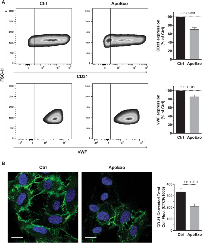
Apoptotic exosome-like vesicles decrease the expression of endothelial markers in endothelial cells. Apoptotic exosome-like vesicles (ApoExo) decreased CD31 and vWF expressions in endothelial cells. (A) CD31 and vWF expression by flow cytometry analysis in serum-starved endothelial cells exposed for 24 h to the vehicle (Ctrl) or apoptotic exosome-like vesicles (ApoExo). Flow cytometry experiments expressed as the percentage of vehicle-treated cells median fluorescence intensity (50,000 events/sample) ± SEM (right). Representative gates of CD31 and vWF expression are depicted (left). n ≥ 5 for each condition. P values obtained by a one-sample t-test. (B) ApoExo decreased CD31 expression in endothelial cells by confocal microscopy. (Scale bar: 20 µm). Semi-quantitative measurement of CD31 expression depicted as corrected total cell fluorescence (CTCF) ± SEM (right). Representative pictures of CD31 expression are presented (left). CTCF represents the integrated density minus the area of selected cell multiplied by the mean fluorescence of background reading. n = 3 for each condition. P value obtained by unpaired t-test.

