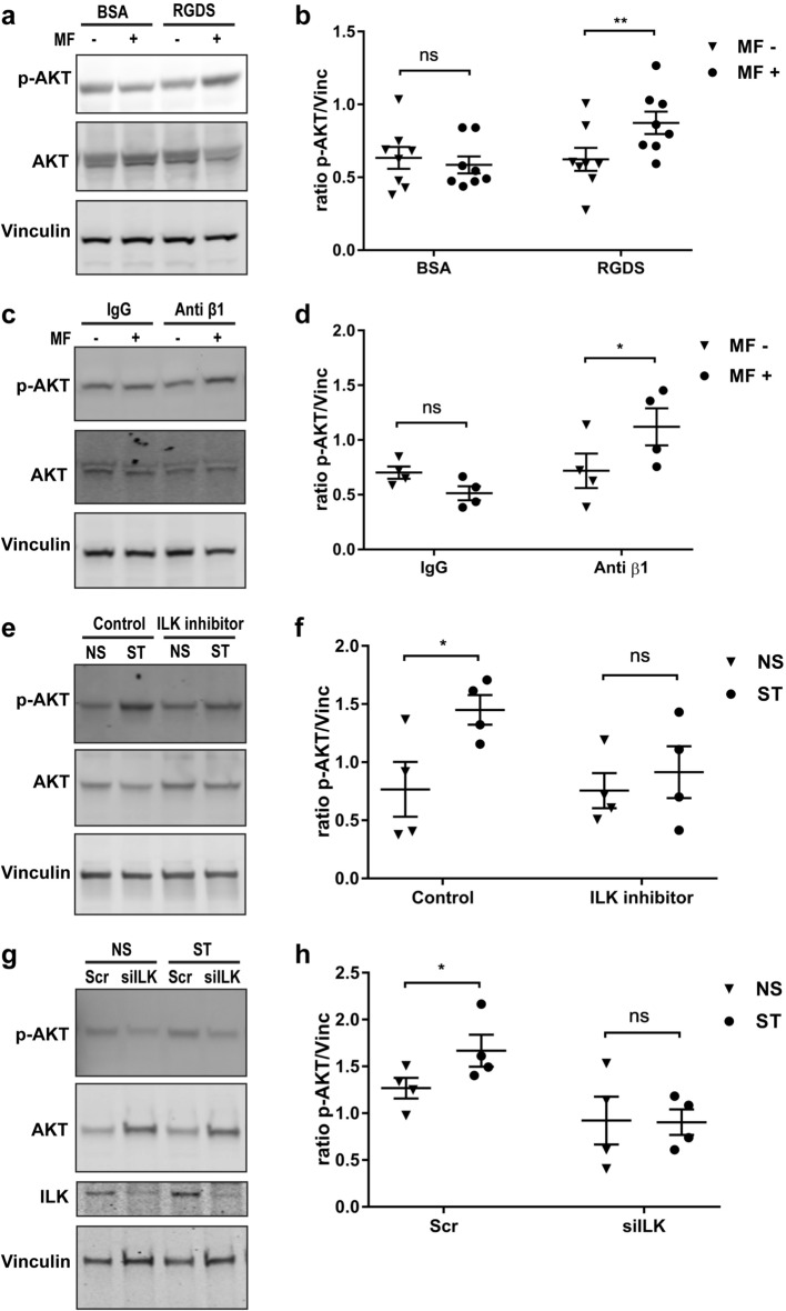
Figure 2.
Mechanical stretching modulates AKT phosphorylation through β1 integrin and ILK. (a) Immunoblot of human tendon cells that were bound to RGDS-conjugated magnetic particles and exposed to one hour of oscillatory magnetic field (MF) with a frequency of 1 Hz. “- “ and “ + ” indicates without or with magnetic field, respectively. (b) shows the densitometry of phospho-AKT normalized to Vinculin in (a). (c) Immunoblot of human tendon cells incubated with anti-integrin β1 conjugated magnetic particles after 1 h of oscillatory magnetic field (MF) with a frequency of 1 Hz. The immunoblots of human tendon cells incubated with ILK inhibitor QLT0267. (d) shows the densitometry of phospho-AKT normalized to Vinculin in (c). (e), and siILK (g) indicate preventing AKT phosphorylation by mechanical stretching (ST) with Flexcell compared to the non-stretched (NS) cells. (f), (g) show the densitometry of phospho-AKT normalized to Vinculin in (e) and (g), respectively. Two-way ANOVA followed by Bonferroni's multiple comparisons test; mean ± SE; ns P > 0.05; * P ≤ 0.05; **P ≤ 0.01; n ≥ 4 biological replicates.

