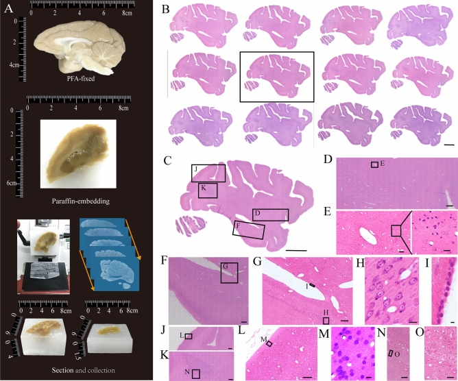
Figure 3.
Slices from an intact macaque hemisphere embedded by paraffin, stained with H&E. (A) Histological sectioning. (B) The serial sagittal plane slices from the macaque hemisphere. All the sections were sectioned at the coronal plane for 5 μm. (C) Magnification of the box in (B). (D) Magnification of the box labeled D in (C). (E) Magnification of the box labeled E in (D). (F) Magnification of the box labeled F in (C). (G) Magnification of the box labeled G in (F). (I, H) Magnification of the boxes labeled I and H in (G). (J, K) Magnification of the boxes labeled J and K in (C). (L) Magnification of the box labeled L in (K). (M) Magnification of the box labeled M in (L). (N) Magnification of the box labeled N in (K). (O) Magnification of the box labeled O in (N). Scale bars, (B, C) 1 cm; (D) 1 mm; (E) 100 μm, black box, 20 μm; (F) 1 mm; (G) 200 μm; (H) 20 μm; (I) 5 μm; (J, K) 1 mm; (L) 200 μm; (M) 10 μm; (N) 200 μm; and (O) 50 μm.

