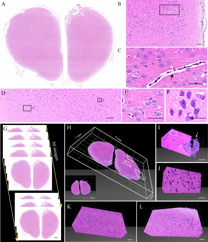
Figure 4.
Slices from an intact rabbit brain embedded by paraffin, stained with H&E. (A) The coronal plane slices from the rabbit brain. (B) Magnification of (A). (C) Magnification of (B). Blood vessels indicated by the black arrow. (D) Magnification of (A). (E, F) Magnification of (D). (G) The coronal plane slices from the rabbit brain. All the sections were sectioned at the coronal plane for 4 μm, and we collected serial 50 images. (H) 3D reconstructed images of (G). (I) Magnification of (H). The meninx indicated by the white arrow. (J) Magnification of (I). (K, L) Magnification of (H). Scale bars, (A) 500 μm; (B, C) 50 μm; (D) 100 μm; (E) 50 μm; (F) 25 μm; (G) 500 μm; (H) white grid, 1 mm; (I) white grid, 100 μm; (J) white grid, 50 μm; (K, L) white grid, 100 μm.

