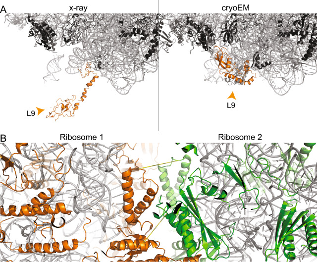
Figure 7.
Multimeric states of the ribosome. Ribosomal proteins (black) and rRNA (grey) are shown in cartoon presentation. (A) Different conformations of L9. The protein is localized in the interface region of the asymmetric unit in the crystal structure (4V4H), and, thereby, is in an extended conformation. In the cryo-EM structure (5U9F), the protein is folding back onto the surface of the ribosome. (B) Cross-linking reveals presence of hibernated ribosomes in the sample (PDB ID 6H58). Ribosomal proteins of the two ribosomes are color-highlighted (green/orange). Cross-links #81 and #97 are shown. The images of protein structures were generated with PyMOL (Version 2.3.2), Schrödinger, LCC. https://pymol.org.

