Figure 3.
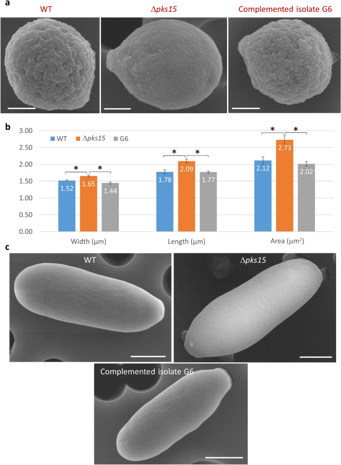
(a) Scanning electron micrographs (SEMs) of conidia from the wild type (WT), Δpks15 and the complemented isolate G6. It is noted that wild-type and G6 conidia have characteristic rodlet bundles on the wall surface but the pks15 mutant lacks these bundles. Also, this Δpks15 conidium, as a representative of most of the mutant conidia, is larger and more elongated than that of the wild type and G6. Bars, 500 nm. (b) Measurement of width, length and area of conidia from the three strains, as analyzed from the electron micrographs taken. Data shown are mean ± S.E.M. Asterisks indicate statistical significance between the wild type or the complemented isolate G6 and Δpks15 (Student’s t test: *p < 0.05). (c) SEMs of blastospores from WT, Δpks15 and G6. Bars, 1 µm.

