Figure 5.
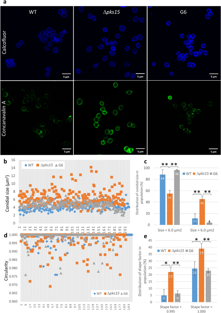
Size and shape of fluorescently-stained conidia in the wild type (WT), Δpks15 and the complemented isolate G6. (a) Calcofluor- and FITC-tagged concanavalin A staining (upper and lower panels). Bars, 5 μm. (b) Distribution of sizes in the conidial populations of the wild type, Δpks15 and G6 from a single representative experiment. (c) Frequency of conidial sizes for each of the three strains. Size data were from three independent experiments. (d) Distribution of shapes in the conidial populations of wild type, Δpks15 and G6 from a single representative experiment. Shape factor (circularity) was determined using the NIS-Elements D software. A shape factor of 1.0 indicates a circle, whereas a shape factor less than 1.0 indicates an ellipse. (e) Frequency of conidial shapes for each of the three strains. Data shown are mean ± s.e.m. Asterisks indicate statistical significance between the wild type or the complemented isolate G6 and Δpks15 (Student’s t-test: *p < 0.05; **< 0.01).

