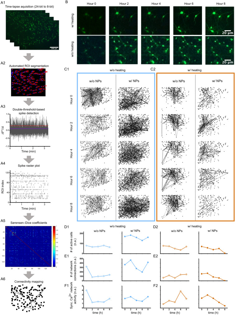
Figure 5.
Chitosan-coated magnetic nanoparticles (NPs) impacts synchronicity in calcium signalling. (A1–A6) Image processing analysis to extract spatially resolved neuronal connectivity map based on primary cortical neurons labelled with Fluo-4. (A1) Time-lapse images show pseudo-coloured calcium activity captured with digital fluorescent microscopy. (A2) Automated image segmentation spatially selects individual regions of interest (ROIs) based on fluorescently active cell bodies. (A3) Changes in calcium activity get recorded over time and (A4) transferred into a calcium spike raster plot based on calcium influx. (A5) A cross-correlation matrix presents high (blue) and low (red) probability of temporally connected calcium spiking activity, which is used in (A6) to connect the ROIs in a connectivity map. (B) Representing fluorescent images of primary neurons incubated for 10 h in the digital imaging system. (C1–C2) Connectivity maps present changes in synchronous calcium spiking activity over an 8-h time span in cortical neurons grown for 9 days in vitro, independent of room temperature (w/o heating), physiological temperature (w/heating) and of incubation with chitosan-coated magnetic NPs (w/NPs). (D1–F2) Network analysis shows (D1–D2) number of active neurons under different temperature conditions, with and without NPs, (E1–E2) number of connections between ROIs under different temperature conditions, with and without NPs, and (F1–F2) active cell normalized count of connections under different temperature conditions, with and without NPs. These four plots indicate the activity of the network and how it changes under the different culture conditions.

