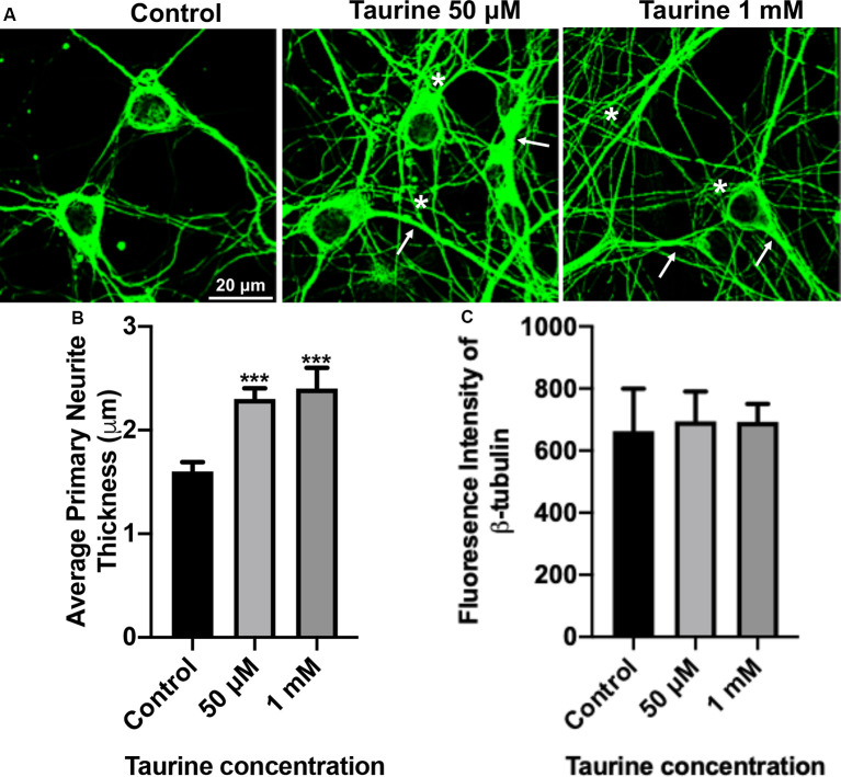
Figure 2.
β-tubulin-labeled cortical neurons reveal an increase in the thickness of neurites after taurine treatment. After 3 days in taurine-rich (50 μM or 1 mM) or -free media, cortical neurons were stained with the cytoskeletal marker β-tubulin and imaged. (A) Representative images demonstrate the extensive outgrowth of cultures in all treatments and control. Arrows indicate examples of extensive neuritic processes while asterisks show robust interconnections. (B) Taurine at both 50 μM and 1 mM concentrations significantly increased the average thickness of primary neurites compared to control. The average neurite thickness for control was 1.6 ± 0.09 μm, for 50 μM taurine was 2.3 ± 0.1 μm, and for 1 mM taurine was 2.4 ± 0.2 μm (p = 0.0004 for control vs. 50 μM taurine, and p = 0.0002 for control vs. 1 mM taurine). (C) The intensity of β-tubulin was not significantly changed by any concentration of taurine. Specifically, the β-tubulin intensity of control was 663.3 ± 136.2, for 50 μM taurine was 694.3 ± 96.3, and for 1 mM taurine was 692.5 ± 58.3 (p = 0.971; n = 4 images per treatment and number of neurites analyzed was 40, 39, and 30 for control, 50 μM taurine, and 1 mM taurine, respectively; the statistical test was one-way ANOVA in all cases). ***Indicates a significance level of p < 0.001 vs. control.

