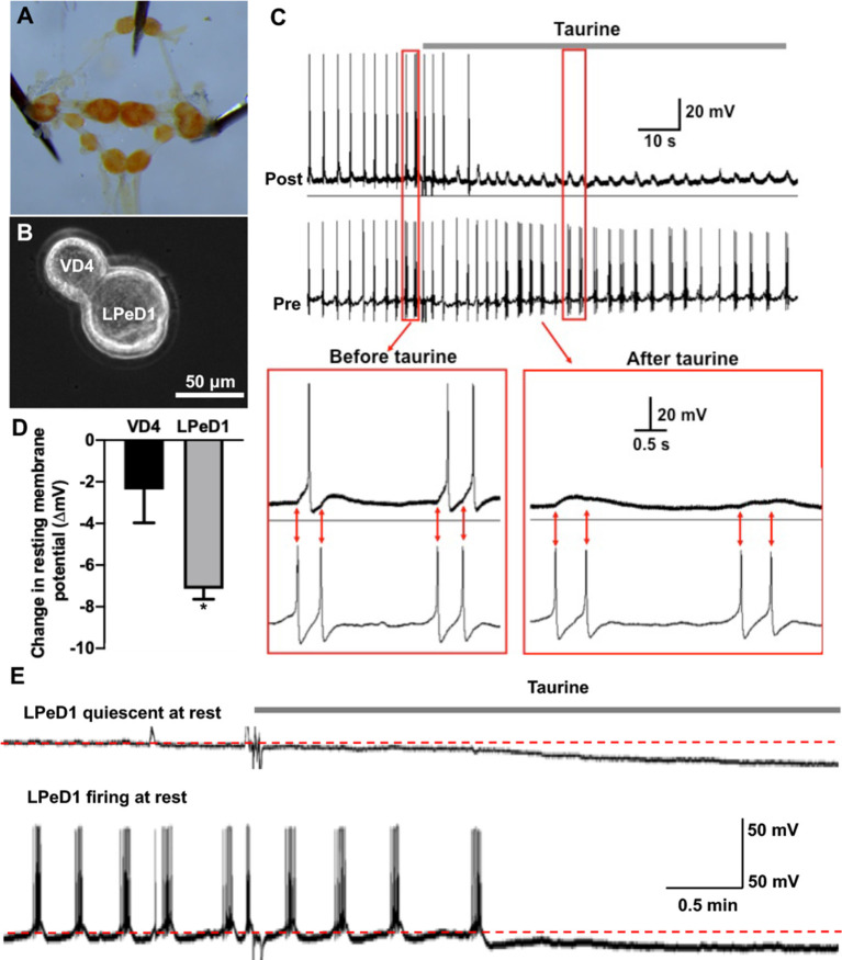
Figure 5.
Acute exposure of Lymnaea central neurons to taurine alters the excitability of two synapse-forming neurons. (A) L. stagnalis central ring ganglion was dissected. (B) To examine whether acute exposure to a high concentration of taurine affects neuronal excitability and/or synaptic transmission, well defined L. stagnalis pre- and postsynaptic VD4-LPeD1 neuronal pairs were cultured and allowed to form synapses (n = 4). (C) Intracellular recordings revealed that presynaptic action potentials elicited electrical activities in postsynaptic neurons, and these were quieted after exposure to taurine at 2.5 mM. Interestingly, the postsynaptic potentials (PSPs) remained throughout the presence of taurine, indicating that taurine can selectively modulate neural excitability change while allowing the synaptic transmission to occur between two synaptic neurons. (D) At rest, taurine caused a significantly larger hyperpolarizing membrane potential change (ΔmV) in LPeD1 neurons (7.14 ± 0.5 mV) than that in VD4 neurons (2.37 ± 1.6 mV; student t-test, p = 0.049; n = 3). Negative direction and Y-axis values indicate a hyperpolarizing action of taurine on the resting membrane potentials of VD4 and LPeD1. (E) The hyperpolarization in LPeD1 neurons occurred in either quiescent or actively firing LPeD1 cells. The dotted lines indicate basal membrane potential levels. *Indicates a significance level of p < 0.05.

