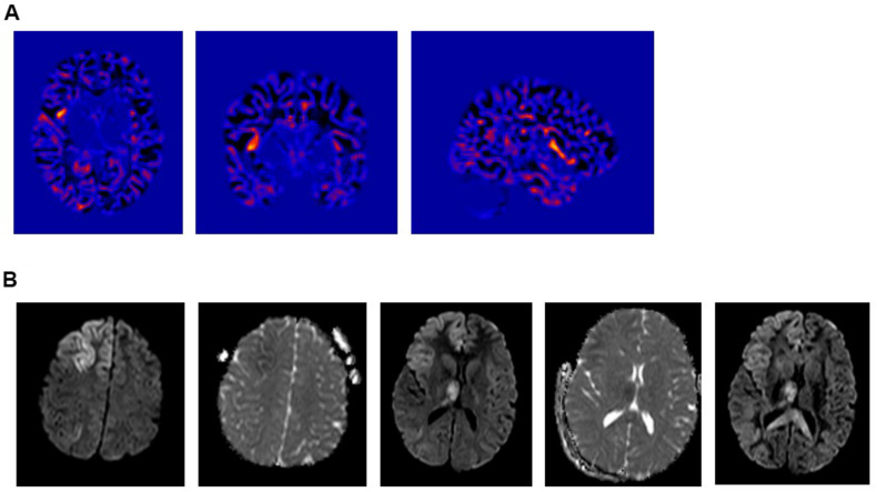
FIGURE 2.
(A) Voxel Based Morphometric (VBM) Analysis – Junction Map. (i) Axial; (ii) Coronal, and (iii) Right Sagittal cuts. Showing a suspicious lesion involving the right anterior insula with a high Z-core. (B) Intra-ictal brain MRI diffusion weighted images (DWI) showing a restricted diffusion in the cortical-subcortical area of the right frontal lobe, right insula and right thalamus.

