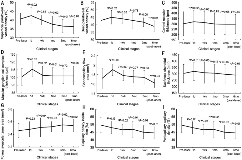
Figure 4. Vascular and thickness changes at different clinical stages.
Superficial and deep parafoveal vessel density (A, B), choriocapillary flow area (E), and subfoveal choroidal thickness (F) were significantly increased at 1d post-laser therapy and decreased continuously to less than baseline level at 6mo post-laser therapy. Central macular thickness (C) and macular ganglion cell complex thickness (D) significantly increased at 1d post-laser therapy and decreased to baseline level at 6mo post-laser therapy. Foveal avascular zone area (G) continuously increased at 6mo after laser therapy. Capillary density inside the disc (H) and peripapillary capillary density (I) continuously decreased at 6mo after laser therapy. The P value indicated that the above parameters at certain stages were (not) significantly different from that at baseline. aP<0.05.

