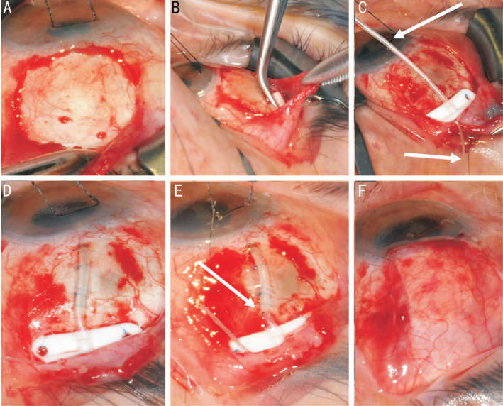
Figure 2. Stages of Baerveldt tube insertion.
A: Subtenons pocket is fashioned in the superotemporal quadrant; B: Mitomycin C can be applied using a PVA corneal shield in the subtenons pocket; C: The place is secured to sclera through the eyelets of the plate (note the supramid suture within the tube, and coming out the end plate); D: The tube is inserted into the anterior chamber fixed with two mattress sutures; E: An external ligating suture is secured; F: The patch graft is sutured over the tube and the conjunctiva and subtenons closed.

