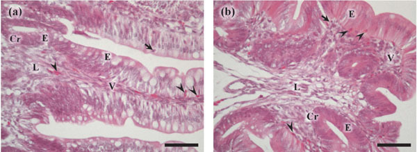
Fig. 1.

Histology of the ileum (a) and cecum (b) of 3-d-old chicks. The villi (V) and crypt (Cr) are lined by luminal and crypt epithelium (E). Eosinophilic heterophil-like cells (arrow heads) are localized in the luminal and crypt epithelium and lamina propria (L) in both the ileum and cecum. Arrows show lymphocytes. Hematoxylin and eosin staining. Scale bars=50 µm.
