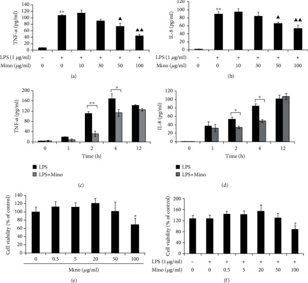
Figure 1.

Minocycline suppressed cytokine and chemokine production in LPS-stimulated THP-1 cells in dose-dependent and time-dependent manner. (a, b) THP-1 cells were stimulated with LPS (1 μg/ml) alone or LPS and (10-100) μg/ml minocycline for 4 h (∗∗p < 0.01 vs. control; ▲p < 0.05, ▲▲p < 0.01 vs. LPS). (c, d) THP-1 cells were stimulated with LPS (1 μg/ml) alone or LPS and 50 μg/ml minocycline for 1, 2, 4, or 12 h (∗p < 0.05; ∗∗p < 0.01). (e, f) The relative cell viability of THP-1 cells stimulated with minocycline or LPS and minocycline for 24 h (∗p < 0.05 vs. control). The concentration of TNF-α or IL-8 in the supernatants was measured by ELISA. Data were represented as the mean ± S.D. (N = 3). Mino: minocycline.
