
Figure 5. Upregulated P53 pathway and DNA damage following prolonged mitosis.
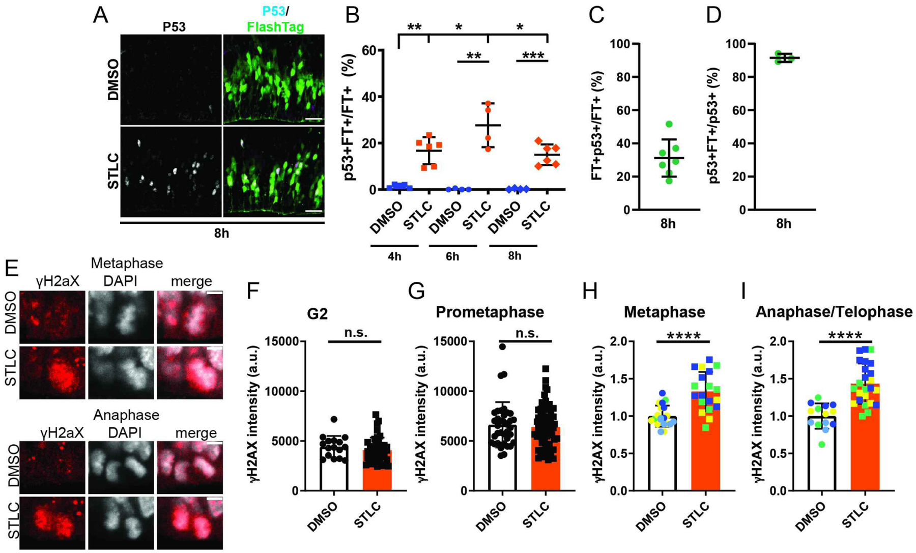
A) Images of the VZ/SVZ depicting FT (green) and p53 (white) 8h after injection of DMSO or STLC. b) Quantification of the fraction of FT+ cells that are CC3+FT+ cells 4h, 6h, and 8 h after STLC injection via analysis of 200micron cortical columns. Dots represent individual embryos, average of 2 or more sections per embryo. 6h**: p=0.004, 8h ****: p<0.0001, 10h ***: p=0.0003, 6h v 8h *:p=0.02, 8h v 10h: p=0.04. C) Quantification of the fraction of FT+ cells that are p53+ at 8h in STLC condition. C) Percent of p53+ cells labeled by FlashTag 8h after STLC injection. N=3 embryos, average of 3 sections per embryo. E) Images of H2AX (red) and DAPI (white) in mitotic cells at the ventricle 3h after injection of DMSO or STLC. F) Quantification of H2AX intensity of mitotic cells at the ventricle 3h after DMSO or STLC injection. Dots represent individual cells across two or more embryos. Note for STLC, primarily FT+ cells were quantified in order to enrich for mitotic cells. Statistics: student’s t-test. metaphase ****: p<0.0001. anaphase/telophase ****: p<0.0001. Scale bar: A, 25 μm, E, 5 μm, Error bar= s.d.