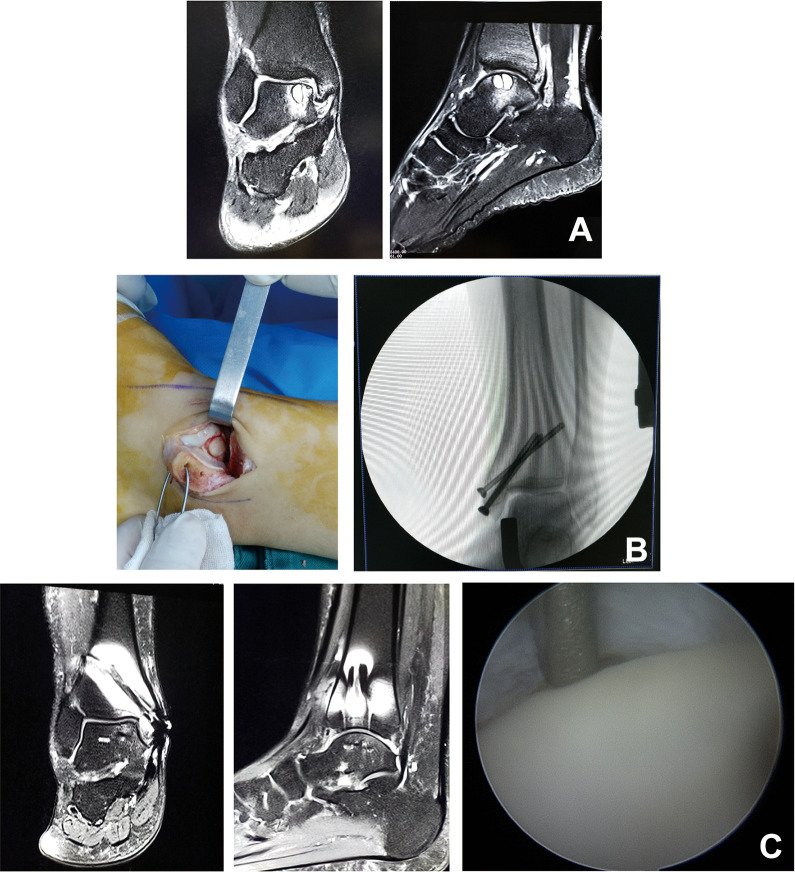
Figure 1.
A 33-year-old male patient. (A) Magnetic resonance imaging (MRI) showed a large talar cyst. (B) Medial malleolar osteotomy was performed for osteochondral grafting and fixed with 3 cannulated screws. (C) At 2 years after surgery, second-look arthroscopic surgery and MRI showed nearly normal cartilage.

