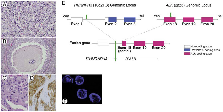
Figure 3.
SDC12 harboring HNRHPH3-ALKfusion formed invasive nests (H&E, medium power, A), showed comedo-type necrosis (H&E, low power, B), apocrine cytology with abundant eosinophilic cytoplasm and prominent nucleoli (H&E, high power, C), and positive cytoplasmic immunostain for ALK (high power, D). Schematic diagrams of HNRNPH3 and ALK partial gene structures as well as the formation of the HNRNPH3-ALK fusion gene are depicted: HNRNPH3 is located at 10q21.3 on the plus strand of chromosome 10, and ALK is located at 2p23 on the minus strand of chromosome 2. Transparent boxes represent noncoding parts of mature transcript of HNRNPH3, blue boxes represent HNRNPH3 coding parts/exons, and red boxes represent ALK coding exons. Green arrows represent break points at HNRNPH3 locus and ALK locus, respectively. The green line represents the fusion point of HNRNPH3-ALK (E). ALK rearrangement was confirmed by FISH (3’ALK, red signal, F). Abbreviations: cen=centromere, tel=telomere.

