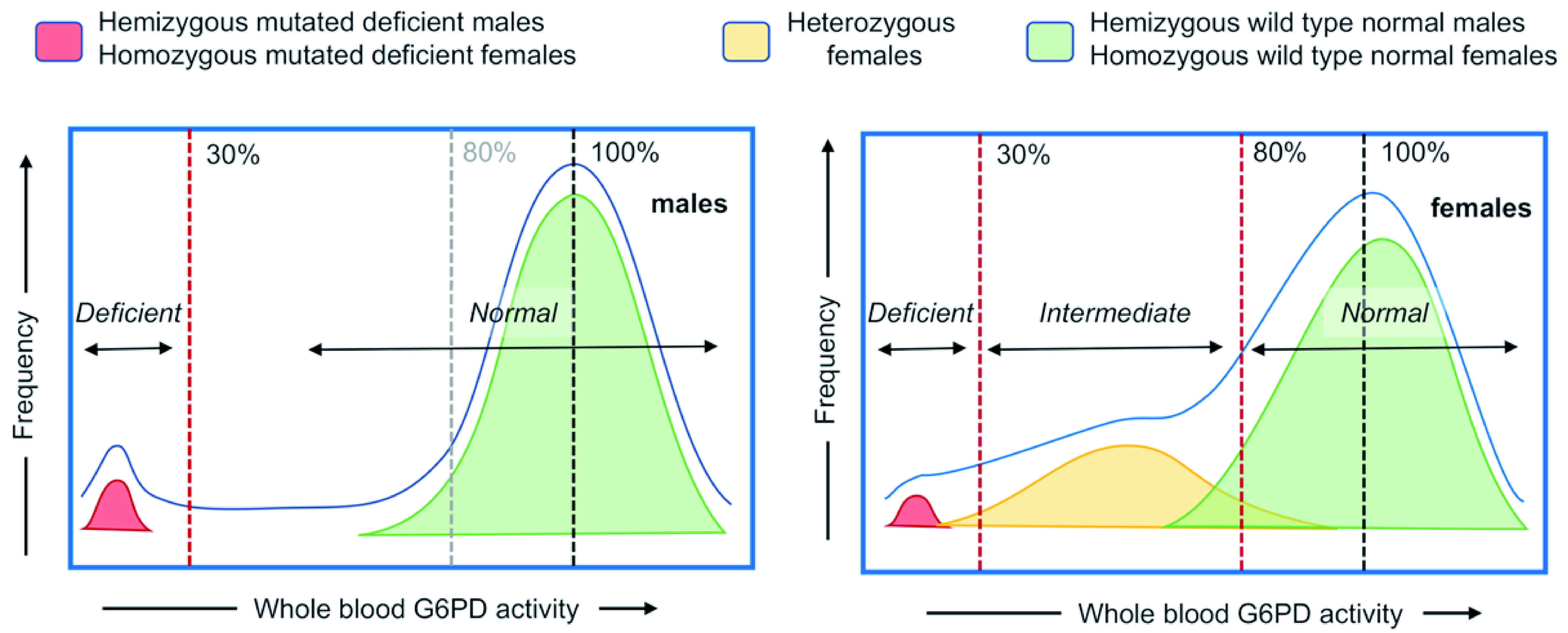
Figure 1. Schematic of population histograms demonstrating the relationship between phenotype and genotype in G6PD deficiency in males (left panel) and females (right panel).

The G6PD gene is located on the X chromosome, such that females have two genes and males have only one. Males with a mutated G6PD allele (in red, G6PD DEF) that expresses a compromised (deficient) G6PD enzyme protein typically have a blood G6PD value of less than 30% of normal. Females with two mutated G6PD-deficient alleles (in red, G6PD DEF1, DEF2) also typically have a blood G6PD value of less than 30% of normal. Males with a wild type G6PD allele (in green, G6PD WT) that expresses a fully functional enzyme have G6PD activity in an approximate normal distribution around the 100% median, as do females with two wild type G6PD alleles (in green, G6PD WT1, WT2). Heterozygous females with both wild type and mutated G6PD alleles (in yellow, G6PD WT, DEF1) can express a spectrum of whole blood G6PD activity, ranging from severely deficient (<30%) to beyond the World Health Organization definition of normal for females (>80%), with the majority in the intermediate (30% to 80%) activity range. The colored zones indicate the distribution of enzymatic activities associated with the genotypes as described above; the blue line represents the cumulative G6PD activity-based histogram.
