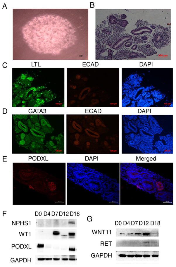
Figure 3.
3D culture of kidney organoids. (A) Bright field observations of the kidney organoids. (B) Hematoxylin and eosin staining results of the kidney organoids. (C) Expression detection of renal tubular markers LTL and ECAD in kidney organoids by immunofluorescence. (D) Expression detection of collection tube markers GATA3 and ECAD in kidney organoids by immunofluorescence. Scale bars, 100 µm. (E) Expression detection of kidney podocytes marker PODXL in kidney organoids by immunofluorescence. Scale bars, 50 µm. (F) Expression detection of the molecular markers of podocyte (NPHS1, PODXL and WT1) by western blotting. (G) Expression detection of the specific markers for ureteric buds RET and WNT11 by western blotting. The data are representative from a minimum of three independent experiments. LTL, Lotus tetragonolobus lectin; ECAD, e-cadherin; GATA3, GATA binding protein 3; NPHS1, nephrin; PODXL, podocalyxin; WT1, Wilms tumor protein; RET, proto-oncogene tyrosine-protein kinase receptor Ret; WNT11, protein Wnt-11; D, day.

