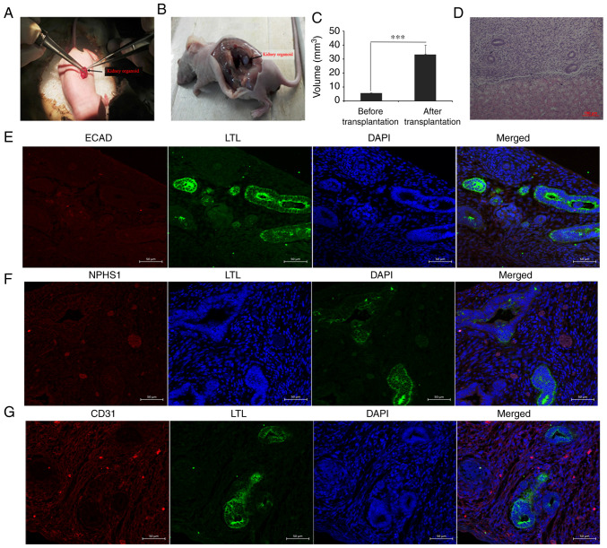
Figure 4.
In vivo cultivation and identification of kidney organoids. (A) Kidney organoids were transplanted into the murine renal capsule, and the graft was removed after 2 weeks of culture in vivo. (B) In vivo images two weeks after transplantation. (C) Statistical analysis before and after transplantation. ***P<0.001. (D) Graft hematoxylin and eosin staining. Scale bar, 100 µm. (E) Expression detection of renal tubular markers LTL and ECAD in the grafts. (F) Immunofluorescence detection of the molecular marker NPHS1 of renal podocytes in grafts. (G) Immunofluorescence detection of vascular endothelial cell molecular marker CD31. Scale bars, 50 µm. LTL, Lotus tetragonolobus lectin; ECAD, e-cadherin; NPHS1, nephrin.

