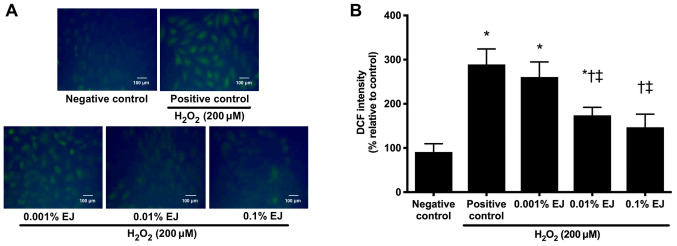
Figure 2.
DCF staining and subsequent confocal fluorescence microscopy findings of 200 µM H2O2-treated human corneal epithelial cells with or without pretreatment with EJ extracts. (A) Representative microscopy images of DCF staining and (B) Relative fluorescence intensity results. Data are expressed as the percentage normalized to the negative control. *P<0.05 vs. negative control; †P<0.05 vs. positive control and ‡P<0.05 vs. the 0.001% EJ group. EJ, Eurya japonica; DCF, dichlorodihydrofluorescein diacetate; H2O2, hydrogen peroxide.

