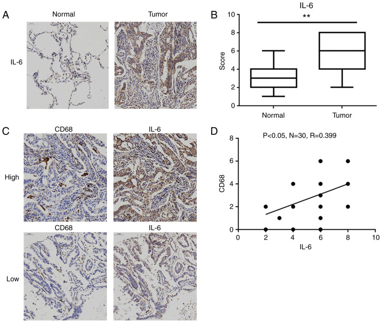
Figure 7.
IL-6 levels are higher in human lung adenocarcinoma tissues and corresponding TAM infiltration. (A) Representative immunohistochemistry images of IL-6 in lung adenocarcinoma tissues and matched normal lung tissues (magnification ×200). (B) Statistical analysis of the immunohistochemistry scores for the expression of IL-6 in 30 pairs of lung adenocarcinoma tissues and matched normal lung tissues, **P<0.01, compared with the matched normal lung tissue group. (C) Representative immunohistochemistry images with high and low levels of IL-6 and CD68 (magnification ×200). (D) Scatter chart showing a positive correlation between the expression levels of IL-6 and CD68 in 30 pairs of lung adenocarcinoma tissues. IL-6, interleukin-6; TAM, tumor-associated macrophage.

