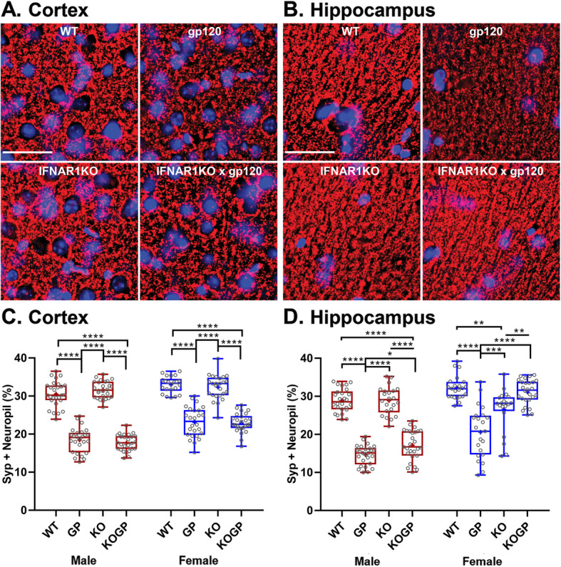
Fig. 2.

IFNAR1 deficiency partially ameliorates the loss of presynaptic terminals preferentially in the hippocampus of females. Representative images of the cortex (a; layer 3) and hippocampus (b; CA1) immunolabeled for neuronal synaptophysin (SYP); deconvolution microscopy; scale bar, 40 μm. c, d Quantification of microscopy data obtained in the cortex and hippocampus of sagittal brain sections of 12–14-month-old mice. Genotypes: WT (WT), HIVgp120tg (GP), IFNAR1KO (KO), and IFNAR1KO × gp120 (KOGP). Values are presented in combined box-dot plots with the 25th and 75th percentiles. The middle line of the box shows the median, and the mean is indicated by a “+”; ****P < 0.0001, ***P < 0.001, **P < 0.01, *P < 0.05; ANOVA and Tukey’s HSD post hoc test; n = 6 animals (3 males and 3 females) per group/genotype (total n = 24 animals)
