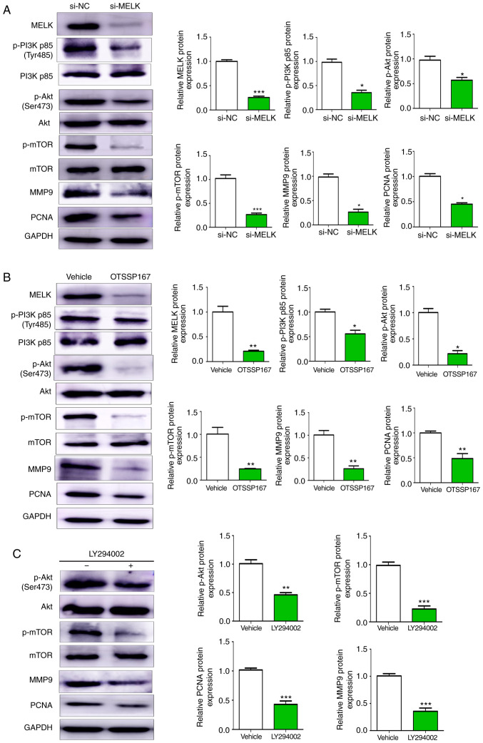
Figure 6.
MELK promotes osteosarcoma proliferation and metastasis through the PI3K/AKT/mTOR pathway. (A) Western blot analysis of changes in protein expression following knockdown of MELK. Cells transfected with si-MELK exhibited lower expression levels of p-PI3K p85 (Tyr485), p-AKT (Ser473), p-mTOR, MMP9 and PCNA. (B) Western blotting revealed lower protein expression levels of p-PI3K, p-AKT, p-mTOR, MMP9 and PCNA in the cells treated with OTSSP167 compared with the vehicle control. (C) PI3K inhibitor LY294002 decreased the expression levels of p-AKT, p-mTOR, PCNA and MMP9. GAPDH was used as the internal control. *P<0.05, **P<0.01, ***P<0.001. MELK, maternal embryonic leucine zipper kinase; si, small interfering; p-, phospho-.

