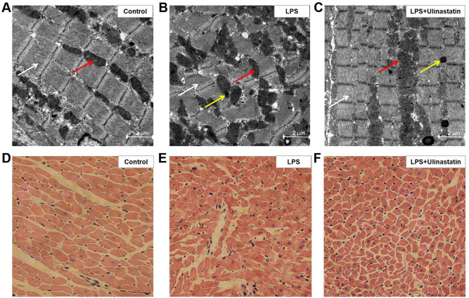
Figure 3.
Effect of ulinastatin on attenuating myocardial injuries. LPS-induced endotoxemia mice were treated with saline or ulinastatin, and the left ventricle tissues were collected 12 h later for analysis. Transmission electron microscopy was used to analyze the cardiac ultrastructure in the (A) control, (B) LPS and (C) LPS + ulinastatin groups. Magnification x15,000. Scale bar, 2 µm. White arrow indicates the myofilaments, the red arrow indicates the mitochondria and the yellow arrow indicates the presence of autophagosomes. Hematoxylin & eosin staining was used to determine the pathological changes in the myocardial tissue in the (D) control, (E) LPS and (F) LPS + ulinastatin groups. Magnification x200. LPS, lipopolysaccharide.

