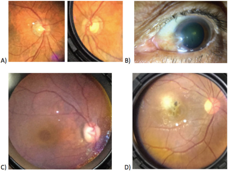
Figure 4:
Examples of ocular pathology as captured on anterior and posterior segment photographs with the Paxos Scope. A) Asymmetrical cupping in fundus photos of the right eye (left side) and left eye (right side) B) Pterygium located on the medial portion of the left eye of the subject C) Macular hole in the fundus photo of a right eye.

