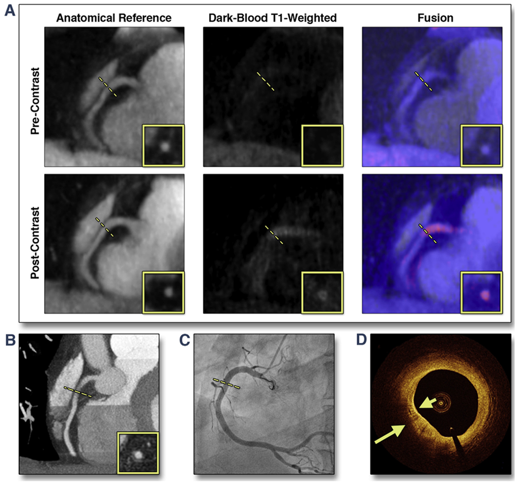
FIGURE 4. Representative Case of a Suspected CAD Patient With a CHIP in the Proximal RCA on Post-Contrast CATCH.

(A) Pre- and post-contrast T1-weighted, anatomical reference and fusion images. (B) Computed tomography angiography. (C) X-ray angiography. (D) Optical coherence tomography cross-sectional image at the corresponding location of the CHIP on CATCH. Arrows point to areas with multiple strong back reflections, suggesting macrophage clusters. Dotted lines represent the location and orientation of the cross-sectional images at the lesion which are shown in boxes. RCA = right coronary artery; other abbreviations as in Figure 3.
