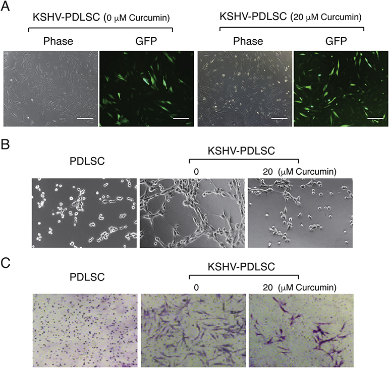
Fig. 4. Effect of Curcumin on KSHV-mediated angiogenesis and cell invasion of MSCs.
(A) Periodontal ligament stem cells (PDLSCs) were infected with KSHV at an MOI of 50 (viral genomic DNA equivalent) in the absence and presence of curcumin as indicated. Infectivity was shown to be 92% and 97%, respectively by GFP expression. (B) Uninfected and KSHV-infected PDLSCs were applied on Matrigel to examine the ability for tubule formation. KSHV-infected PDLSCs were placed on Matrigel in the presence of 20 μM curcumin. Tubulogenesis was examined under a microscope and quantified by measuring the total tube length using the Image J software. (C) PDLSCs or KSHV-PDLSCs (1.5 × 10⁴ cells/well) were seeded in the upper chamber of Transwell with a layer of Matrigel. Cells migrated to the lower chamber were stained with crystal violet. KSHV-infected PDLSCs were treated with 20 μM curcumin and effect of curcumin on cell invasion were assessed by counting migrated cells in the lower chamber. Quantification of transwell-invasion assay was performed by Image J software by counting from multiple randomly selected microscopic visual fields.

