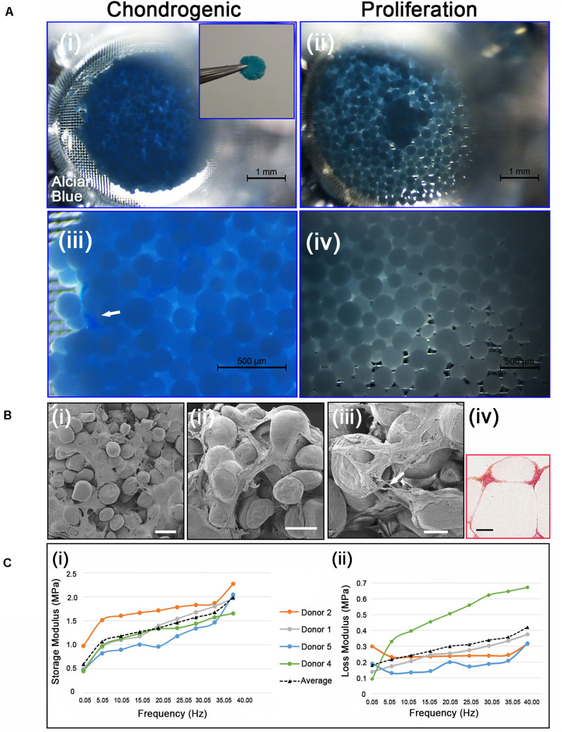
FIGURE 5.
(Ai,iii) Chondrogenic differentiation of cAdMSC on PLGA TIPS microcarriers incubated for 21 days in chondrogenic differentiation medium or (ii,iv) proliferation medium and stained with Alcian blue (Inset shows the disc structure consisting of cellularized PLGA TIPS microcarriers after 21 days incubation in chondrogenic differentiation medium). (Bi–iii) SEM of the disc structure consisting of cellularized PLGA TIPS microcarriers after 21 days incubation in chondrogenic differentiation medium (scale bar 100 μm). Arrow in panel (B) (iii) shows a cell bridging two PLGA TIPS microcarriers; (iv) Picrosirius red staining of collagen in the disc-like structure consisting of cellularized PLGA TIPS microcarriers after 21 days incubation in chondrogenic differentiation medium (red: collagen fibers, yellow: cytoplasm; scale bar 50 μm). (C) (i) Storage modulus and (ii) loss modulus of the disc-like structure consisting of cellularized PLGA TIPS microcarriers after 21 days incubation in chondrogenic differentiation medium measured by dynamic mechanical analysis in compression mode.

