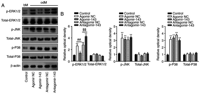
Figure 4.
miR-143 negatively regulated the ERK1/2 signaling pathway. hADSCs were transfected with agomir-143, antagomir-143 or the corresponding NC (100 nM) for 16 h. The medium was replaced with osteogenic medium, and hADSCs were continuously cultured for 14 days. (A) Western blot analysis was used to assess the protein expression levels of p-ERK1/2, ERK1/2, p-JNK, JNK, p-P38 and P38, with β-actin used as an internal control. (B) The bands were semi-quantitatively analyzed using ImageJ software and normalized to β-actin density. Data represent the mean ± standard deviation of three independent experiments. *P<0.05 and **P<0.01, vs. control group; ##P<0.01 and $$P<0.01. miR, microRNA; hADSCs, human adipose-derived mesenchymal stem cells; NC, negative control; ERK1/2, extracellular-signal regulated kinase 1/2; JNK, c-Jun N-terminal kinase.

