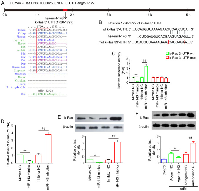
Figure 6.
k-Ras was a direct target of miR-143. (A) Sequences of miR-143 binding sites are highly conserved among different species. (B) Putative binding site of miR-143 and k-Ras. (C) Luciferase activity of hADSCs co-transfected with luciferase reporter constructs containing WT or Mut k-Ras 3′-UTR, and with miR-143 mimics, miR-143 inhibitor or NC (n=3). (D) Reverse transcription-quantitative polymerase chain reaction and (E) western blot analysis of mRNA and protein expression levels of k-Ras in hADSCs transfected with miR-143 mimics, miR-143 inhibitor or NC miRNA (n=3). (F) Western blot analysis of k-Ras protein expression in hADSCs transfected with agomir-143, antagomir-143 and the corresponding NC. The medium was replaced with odM after 16 h, and hADSCs were continuously cultured for 14 days prior to protein level assessment. Data represent the mean ± standard deviation of three independent experiments. **P<0.01 and ##P<0.01. miR, microRNA; hADSCs, human adipose-derived mesenchymal stem cells; WT, wild-type; Mut, mutant; NC, negative control; odM, osteogenic differentiation medium.

