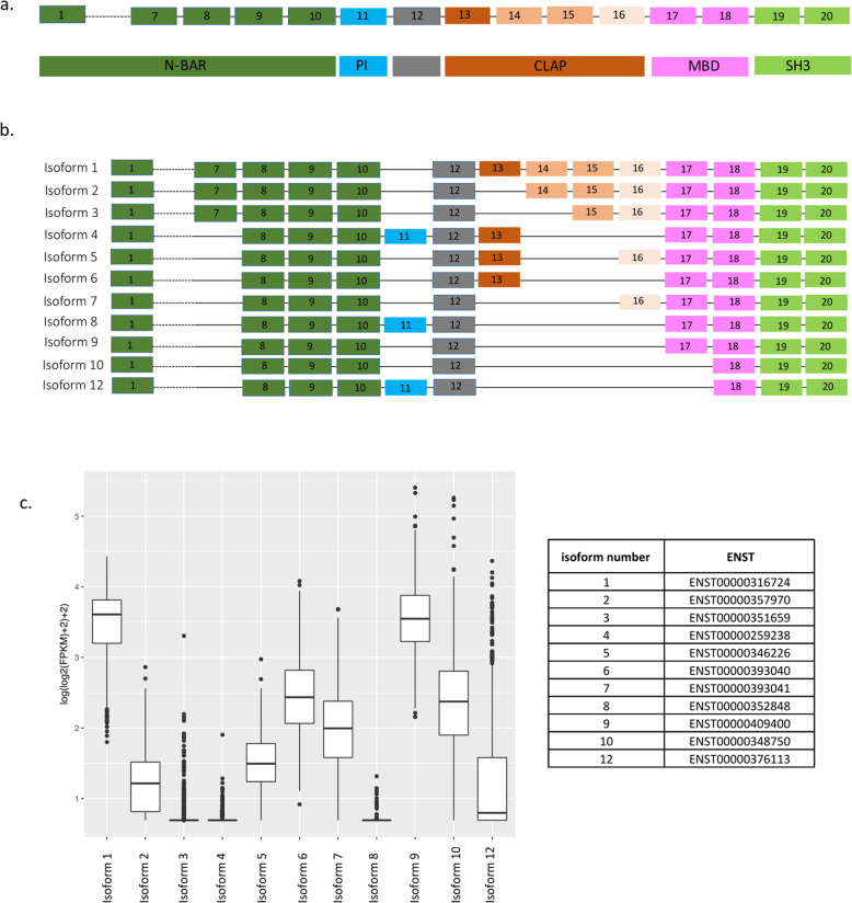
Fig. 1.
BIN1 Isoforms. a The upper aspect of the panel shows the exonic structure of BIN1 along the chromosome, with each exon numbered. Given space constraints, we do not show exons 2–6. The lower aspect of the panel highlights the different domains of the BIN1 protein. Both aspects of the panel are colored based on the functional domains. Glossary: N-BAR domain, phosphoinositide binding module (PI), clathrin and AP2 binding domain (CLAP), Myc-binding domain (MBD), and src homology 3 domain (SH3). b Diagrams of the 12 BIN1 RNA isoforms. c mRNA expression of BIN1 isoforms in human dorsolateral prefrontal cortex (DLPFC) (n = 508 subjects) [6]. These are FPKM values from RNA-seq data, unadjusted for covariates (see Methods section); the data were obtained from subjects with pathological AD (58%) and subjects without AD pathology (42%), and the average age death of 88. We also provide a small table with the ENST reference number for each of the RNA isoforms considered in this study

