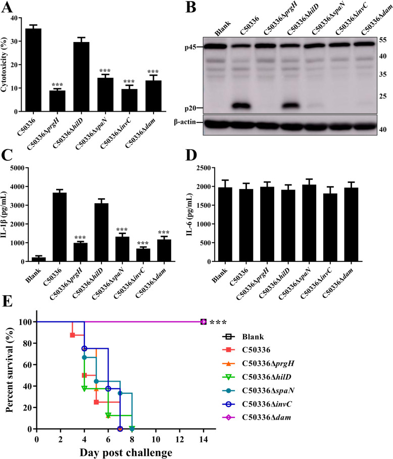
Fig. 2.
Deletion mutants of dam, invC, prgH, and spaN failed to induce inflammasome activation. J774A.1 cells were pre-treated with LPS (1 μg/mL, 5 h) and then infected with WT strain C50336 and dam, invC, hilD, prgH, and spaN gene deletion mutants at an MOI of 20 for 4 h, uninfected cells was used as a negative control (Blank). a. The ratio of cell death was evaluated by the release of LDH in supernatants of infected cells. b. The activation of caspase-1 (p20) was examined via western blot. β-actin was blotted as a loading control. Molecular mass markers in kDa are indicated on the right. Original images of immunoblotting were shown in Fig. S3. The production of IL-1β c and IL-6 d in supernatants were examined via ELISA. ***p < 0.001 for one-way ANOVA followed by Bonferroni’s multiple comparison test indicate significant findings in comparison with cells infected with WT strain C50336. Data are presented as mean ± SEM of triplicate samples per experimental condition from three independent experiments. e. C57BL/6 mice were intraperitoneally injected with PBS (negative control, Blank), C50336, and gene deletion mutants at a dose of 1 × 10⁵ CFU per mouse. The mortality was recorded over 14 dpc. ***, p < 0.001 compared with the C50336-infected group by log-rank (Mantel-Cox) test for the survival curve

