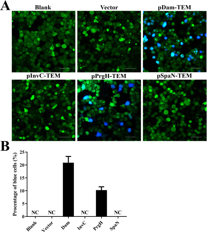
Fig. 3.
Target proteins Dam and PrgH were able to be translocated into the infected cells. HeLa cells were infected with WT C50336 bearing empty plasmid pCX340 or expressing different TEM-1 fusion proteins at an MOI of 100, uninfected HeLa cells was used as a negative control (Blank). Cells were washed and loaded with CCF2-AM after infection. a. Translocation of TEM-1 fusion proteins into the cell cytosol results in cleavage of CCF2-AM, emission of blue fluorescence revealed the activity of TEM β-lactamase, whereas uncleaved CCF2-AM emitted green fluorescence. Scale bar, 50 μm. b. The percentages of cells emitting blue fluorescence. For a particular cell well, six pictures were taken and approximately 1200–2000 cells were counted. Each picture was considered an independent observation and used to calculate the percentage of blue fluorescent cells. Data are presented as mean ± SEM of triplicate samples per experimental condition from three independent experiments

