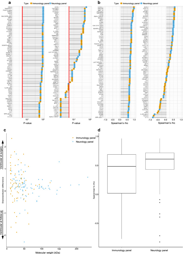
Fig. 4.
Comparison of proteins detected between ventricular CSF at surgery and ventricular CSF obtained at follow-up (between 12 and 60 months post-surgery). a p-values listed for all detected proteins with 0.05 indicated by red line. b Spearman’s rho showing correlation for all detected proteins with lines starting at zero, indicating positive and negative correlation by right and left, respectively. a, b Circles in orange mark proteins from the immunological panel, and circles in blue mark proteins from the neurological panel. Molecular weights (kDa) shown by numbers within coloured circles. c Spread of proteins between compared compartments based on molecular weight (kDa), where a positive standardized difference indicates levels at surgery and a negative standardized difference indicates levels at follow-up. d Boxplot for spread of spearman’s rho subdivided according to the two protein panels

