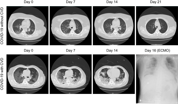
Figure 1.
Representative chest computed tomography (CT) images of COVID-19 pneumonia in a non-cardiovascular disease (CVD) case and a CVD case. Top panel: A 60-year-old man with COVID-19, but not CVD: chest CT images showed ground-glass opacity (GGO) and patchy consolidation with peripheral and subpleural distribution, which had been absorbed at 21 days after hospitalization with treatment. Bottom panel: A 65-year-old man with both COVID19 and CVD: chest CT images showed diffusely subpleural consolidation with a crazy-paving pattern. Diffuse shadowing and consolidation were seen on chest radiography after intensive care unit (ICU) admission with extracorporeal membrane oxygenation (ECMO) support at day 16.

