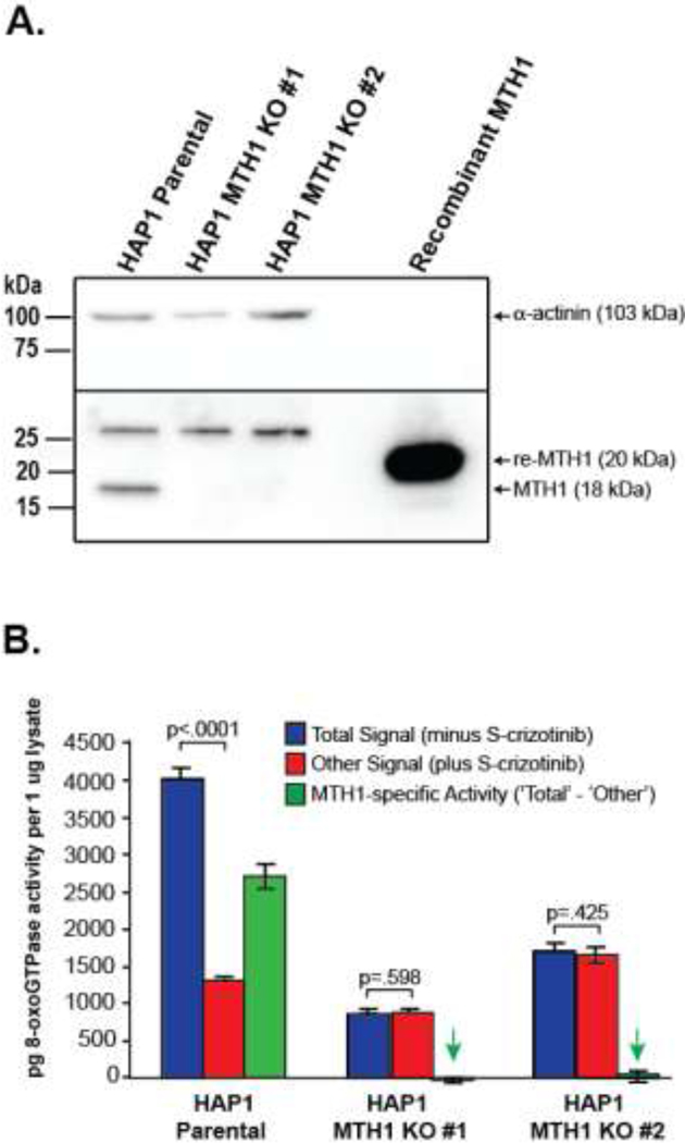
Fig. 2. The ARGO assay measures MTH1-specific 8-oxodGTPase activity.
A. Western blot of MTH1in Hap1 parental cells and two MTH1 KO cell lines demonstrates that there is no MTH1 protein expression in the KO cell lines. Recombinant MTH1 and α-actinin were used as positive and internal controls, respectively. B. ARGO assay showing Total signal (minus S-crizotinib), Other signal (plus S-crizotinib) and MTH1-specific 8-oxodGTPase activity (‘Total’– ‘Other’). No significant differences between ‘Total signal’ and ‘Other signal’ in the MTH1 KO cell lines indicate that in the absence of MTH1 expression, no MTH1-specific 8-oxodGTPase activity is detected by the ARGO assay. Green arrows highlight that there is no significant MTH1-specific 8-oxodGTPase activity in the MTH1 KO cell lines (p-values of .532 and .292 respectively, when compared by two-tailed Student’s T-test to zero).

