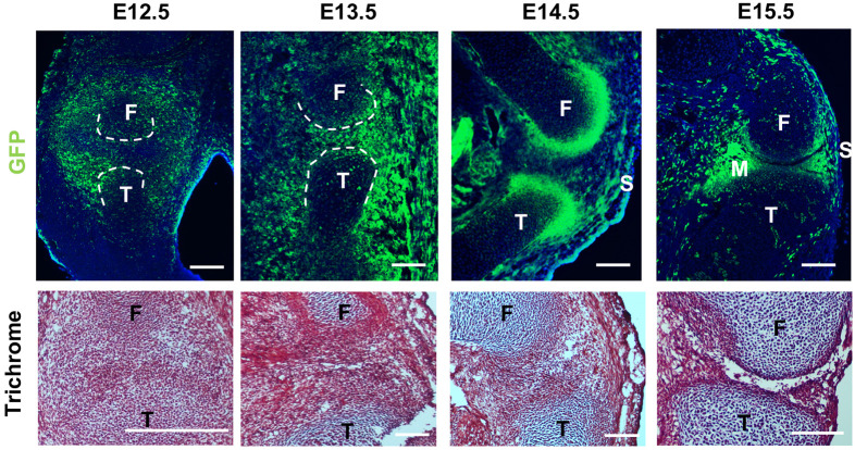
Fig. 1.
Morphology of the embryonic knee joint and localization of Gdf5-lineage cells. Top: localization of Gdf5-lineage cells in murine hindlimb. Bottom: cell density and morphology during joint formation, as shown by trichrome staining. n=6 per timepoint. F, femur; M, meniscus; S, synovium; T, tibia. Scale bars: 100 µm.

