Fig. 3.
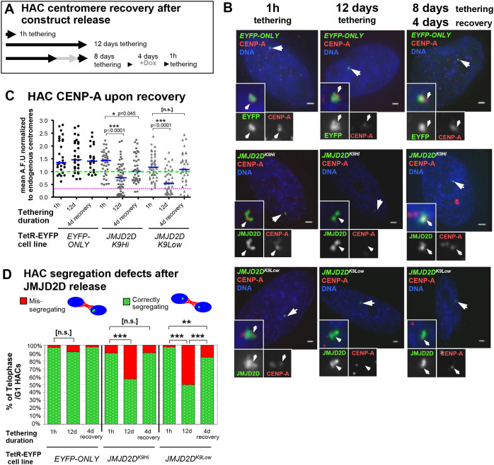
Release of JMJD2D allows for recovery of HAC centromere proteins and mitotic segregation. (A) Outline of the tethering and release strategy, to test centromere recovery. Doxycycline was washed out of cell medium and cells were allowed to grow for 8 days; a fraction of these were allowed to grow for 4 more days, while another had doxycycline added to the medium to prevent JMJD2D binding, for 4 more days. Doxycycline was then washed from the medium to allow TetR-fusion proteins to tether for 1 h only, to allow HAC visualization, before fixation for immunofluorescence. (B,C) Images and quantification of HAC CENP-A recovery after JMJD2D release (see A), in the HAC cell lines expressing the TetR–EYFP fusion proteins. Arrowheads denote the HAC. Scale bars: 2 μm. Data are from two biological repeats, n=14–33 interphase cells each. Blue bar indicates median, green dotted line indicates median starting levels of control EYFP-only HAC CENP-A, magenta dotted line indicates 32.9% of the median endogenous CENP-A level. *P<0.05; ***P<0.0005; n.s., not significant (Mann–Whitney U test). (D) Quantification of HAC segregation defects after JMJD2D release (see A), in the HAC cell lines expressing the TetR–EYFP fusion proteins. Data are from two biological repeats, total of n=20–59 telophase or early G1 cells. **P<0.005; ***P<0.0005; n.s., not significant (Fisher's exact test).

